A jaw lesion can be a fluid-filled cyst, a solid tumor, or a more aggressive growth, and pain cannot tell you which one it is. By the end, you will know which warning signs need prompt assessment, how diagnosis is confirmed, what surgery can prevent, and which questions to ask before treatment.
Key takeaways
- A tumor is not automatically cancer; biopsy identifies the tissue type.
- Panoramic X-rays, CBCT scans and biopsy guide the treatment choice.
- Small, well-defined lesions may need less extensive surgery.
- Ask about recurrence risk, nerve effects, reconstruction and follow-up scans.
Cyst or tumor: what could be growing in the jaw?
A jaw cyst is usually a fluid-filled cavity; a tumor is a solid tissue growth. “Tumor” does not automatically mean cancer: it describes a mass, which may be benign, locally aggressive or, less commonly, malignant. Imaging and tissue examination—not pain alone—determine what is present.
Seek prompt assessment for:
- An enlarging jaw swelling or facial asymmetry
- Loose or displaced teeth, delayed eruption or an altered bite
- Numbness of the lower lip or chin
- Drainage or recurrent infection
- Difficulty opening the mouth
- Trouble swallowing or breathing
A painless lesion still needs evaluation because some cysts grow silently, thinning bone before they cause discomfort. Pain cannot distinguish a cyst from a tumor: infection can make a cyst painful, while a growing tumor can remain painless. Delaying assessment can allow expansion, tooth damage, sinus involvement or, rarely, a pathologic fracture.
| Lesion | What it is often associated with | Why treatment differs |
|---|---|---|
| Radicular cyst | Infection at a tooth root | Treating the source tooth, with cyst removal when indicated |
| Dentigerous cyst | An unerupted or impacted tooth | Treatment may involve preserving, exposing or removing the tooth |
| Odontogenic keratocyst | A cyst with meaningful recurrence risk | Requires careful removal and prolonged imaging follow-up |
| Ameloblastoma | A benign but locally aggressive solid tumor | Wider surgery may be needed to limit bone destruction and recurrence |
The jaw tumor removal benefits depend on the confirmed diagnosis, location and growth risk, not on the label alone.
How doctors identify the lesion before choosing surgery
The diagnosis starts with an oral and facial examination, not with surgery. The clinician checks the swelling, teeth, bite, jaw movement, sensation and drainage, then reviews dental X-rays. A periapical film shows a specific tooth and its roots; a panoramic radiograph surveys both jaws, impacted teeth and the lesion’s broad position.
- Use cone-beam CT to measure the lesion in three dimensions, show cortical expansion or perforation, identify displaced or resorbed teeth, and map the inferior alveolar nerve, maxillary sinus or nasal cavity.
- Use medical CT when the lesion is extensive, reaches the orbit or other facial structures, or requires broader assessment of bone and soft-tissue involvement.
- Use MRI when the key question concerns soft tissue, marrow, muscle or possible nerve involvement. MRI complements CT; it does not replace CT for most bony surgical planning.
| Scan | Best information | Main planning use |
|---|---|---|
| Dental X-ray or panoramic radiograph | Tooth roots, impacted teeth and overall location | Initial assessment |
| Cone-beam CT | Detailed three-dimensional bone and tooth anatomy | Localised jaw surgery planning |
| Medical CT | Larger field and complex facial anatomy | Extensive or sinus-, nasal cavity- or orbit-adjacent lesions |
| MRI | Soft tissue and marrow detail | Selected atypical or soft-tissue questions |
Aspiration involves withdrawing a sample with a needle. Fluid, blood or pus can redirect the diagnosis and reduce the chance of treating the wrong lesion before jaw cyst surgery in Mumbai. Imaging rarely proves the exact pathology.
A biopsy or surgical specimen needs microscopic examination by a pathologist for solid, recurrent, unusually large or atypical lesions.
When surgery helps, and which procedure may be proportionate
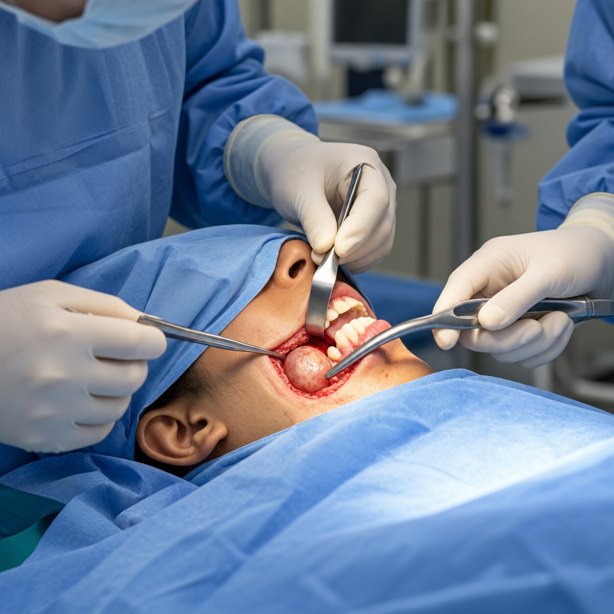
The benefits of jaw cyst tumor surgery in Mumbai are not limited to removing a visible lump. Treating an enlarging lesion can stop bone thinning or perforation, tooth displacement or resorption, infection, maxillary sinus involvement and pathologic fracture.
A small, symptom-free radicular cyst or dentigerous cyst may instead be watched with scheduled X-rays or CT scans when its growth risk is low.
| Option | What it means | When it may fit |
|---|---|---|
| Observation | Imaging at planned intervals | Small, stable radicular cyst with low growth risk |
| Enucleation | Removing the cyst lining intact | Localised dentigerous cyst or radicular cyst when complete removal is practical |
| Curettage | Scraping residual lining from bone | Selected cysts; incomplete removal increases recurrence risk |
| Peripheral ostectomy | Removing a thin rim of surrounding bone | Odontogenic keratocyst, whose recurrence risk can remain significant |
| Marsupialisation or decompression | Creating or maintaining an opening or drain | Large dentigerous cyst or odontogenic keratocyst needing shrinkage before definitive surgery |
Decompression can preserve adjacent teeth and reduce bone removal, but the opening must remain patent. You need regular irrigation, prolonged imaging follow-up and sometimes a second operation for enucleation.
A lesion alone can be removed when nearby roots remain healthy. Root-canal treatment may be needed for a tooth whose pulp is infected; extraction is considered when the tooth is non-restorable, severely resorbed or prevents complete removal. An aggressive ameloblastoma, recurrent odontogenic keratocyst or confirmed malignancy may require wider jaw resection, sometimes with reconstruction.
A pathology report determines whether the planned operation was proportionate.
What to expect after jaw cyst or tumor removal
Pain, swelling, bruising, restricted mouth opening and difficulty chewing are common during early recovery after jaw cyst or tumor removal. Soft foods, prescribed pain relief and careful oral hygiene help, but infection, bleeding, adjacent-tooth damage, jaw fracture and persistent sensory change need prompt review.
Lower-jaw surgery requires a specific discussion of the inferior alveolar and mental nerves. Injury can cause temporary or permanent numbness, tingling or altered sensation in the lower lip, chin or teeth; risk rises when the lesion touches the nerve or removal is extensive.
Upper-jaw surgery instead raises concerns about the maxillary sinus, nasal cavity, orbit and dental roots. An oroantral communication can cause fluid or food to pass between the mouth and sinus.
Tooth preservation is possible, not guaranteed. Recurrence depends on the pathology, completeness of removal and treatment method. Odontogenic keratocysts, for example, need prolonged radiographic follow-up. Histopathology can also change the plan: an apparently routine cyst specimen may reveal an ameloblastoma or another aggressive tumor requiring wider treatment, reconstruction or longer surveillance.
Before discharge or follow-up, confirm:
- Whether the specimen will undergo microscopic histopathology
- Which teeth were preserved, treated or removed
- How to report fever, increasing swelling, bleeding or sinus symptoms
- Whether nerve symptoms are expected and when they need review
- The written imaging schedule and duration of surveillance
A search for jaw cyst treatment Borivali is only a starting point; the pathology report and follow-up plan matter more.
How to compare surgeons and treatment plans in Mumbai
A sound comparison starts with the diagnosis and plan on paper, not the clinic’s location. A search for jaw cyst treatment Borivali or jaw cyst surgery Mumbai should lead you to ask:
- What is the provisional diagnosis, and which scan supports it: dental X-ray, panoramic radiograph, cone-beam CT, medical CT or MRI?
- Is aspiration needed to check for fluid, blood or infection?
- Will all removed tissue be sent for histopathology?
- Why is this procedure preferable to observation, decompression or a more extensive operation?
- What are the nerve, tooth-preservation, reconstruction and tooth-replacement risks?
- How long will pain, swelling, restricted opening and chewing difficulty last?
- Can I receive a written imaging follow-up schedule?
| Option | Main trade-off | Question to ask |
|---|---|---|
| Observation | Avoids surgery but requires scheduled scans | What growth or recurrence would trigger treatment? |
| Decompression or marsupialisation | Preserves bone and nearby teeth but needs irrigation and monitoring | Will a drain or opening remain patent, and could a second operation be needed? |
| Enucleation or curettage | Removes the lesion with less jaw removal | How does the suspected pathology affect recurrence risk? |
| Wider resection | Better control for aggressive disease but may require reconstruction | What reconstruction and tooth replacement will follow? |
The oral maxillofacial surgeon Mumbai you choose should explain functional effects, not only cosmetic results. A large, recurrent, nerve-adjacent or potentially aggressive lesion may need an oral and maxillofacial surgeon, oral pathologist, radiologist, dentist and restorative specialist working together.
Dr. Karan Mehta's Mouth Jaw Face & Hair Transplant may be relevant for assessment in Borivali West, but documented diagnosis, proportionate treatment, pathology review and follow-up matter more than a marketing phrase or address.
Related services
Jaw Jaw Cyst and Tumors Various cysts and tumors can arise in both upper jaw bone(maxilla) and lower jaw bone(mandible). View service → |
Face A cyst is a water-filled cavity. The most common cyst in the oral region is a Radicular cyst arising at the root of teeth due to infection in it. View service → |
Frequently asked questions
What is the difference between a jaw cyst and a jaw tumor?
A jaw cyst is usually a fluid-filled cavity, while a tumor is a solid tissue growth. A tumor may be benign, locally aggressive or malignant, so the name alone does not establish cancer.
How do doctors identify a jaw cyst or tumor before surgery?
Doctors combine an examination with imaging such as a panoramic X-ray or CBCT scan. They use tissue examination, usually through a biopsy or the removed specimen, to identify the lesion.
When can surgery help a jaw cyst or tumor?
Surgery can remove the lesion, obtain a definitive diagnosis, protect nearby teeth and nerves, and reduce the risk of continued bone damage. The appropriate procedure depends on the lesion’s size, location, behaviour and biopsy result.
What should you expect after jaw cyst or tumor removal?
Expect swelling, discomfort and a soft-food period after surgery. Your surgeon will explain wound care, warning signs, pathology results, follow-up visits and whether repeat imaging or reconstruction is needed.
How should you compare jaw surgeons in Mumbai?
Check the surgeon’s oral and maxillofacial training, experience with the specific lesion, planned procedure, pathology arrangements, nerve-risk discussion, reconstruction options and follow-up schedule.
Keywords