A jaw lesion that looks like a cyst on an X-ray still needs a careful diagnosis, a procedure matched to its location, and laboratory confirmation after removal. You will learn which warning signs need urgent review, how the main operations differ, what recovery should look like, and which follow-up questions protect your teeth, nerves, and long-term health.
Key takeaways
- Seek urgent care for breathing difficulty, fever, pus, or uncontrolled bleeding.
- Ask how the cyst’s size and location change surgical risks.
- Follow wound-care instructions and monitor swelling during the first week.
- Request pathology results and attend follow-up even after healing.
When a suspected oral cyst needs urgent assessment
Rapidly increasing jaw swelling, fever, pus, difficulty swallowing or breathing, uncontrolled bleeding, or new facial numbness needs prompt assessment. Fluid passing between the mouth and nose, worsening sinus symptoms, or swelling near the eye also requires urgent review, especially after an upper-jaw lesion has opened into the maxillary sinus.
Seek emergency care for breathing difficulty, spreading neck swelling, confusion, or inability to swallow saliva. For anyone researching oral cyst removal for risks borivali, do not assume a painless lump is harmless; jaw lesions can grow silently and weaken bone.
A surgeon confirms the diagnosis in stages, not from an X-ray alone. The examination checks tooth vitality, mobility, gum swelling, nerve sensation, and whether the lesion is attached to a tooth.
A periapical or panoramic X-ray shows its relationship to roots; CBCT adds three-dimensional detail when the lesion is large or close to the inferior alveolar nerve, sinus, or nasal cavity.
- Ask whether the lesion needs aspiration or an incisional biopsy before definitive surgery.
- Confirm that the entire removed lining will be sent for histopathology.
- Ask how the plan changes if pathology identifies a tumour, infection, or another jaw disease.
| Test | What it shows | Why it matters |
|---|---|---|
| Clinical examination | Tooth, gum, nerve, and swelling findings | Identifies urgent infection and tooth involvement |
| Periapical or panoramic X-ray | Lesion borders and root relationship | Guides the initial diagnosis |
| CBCT scan | Three-dimensional position and bone thickness | Maps nerves, sinus, nasal cavity, and fracture risk |
| Histopathology | Microscopic identity of the lining | Confirms whether it is truly a cyst after removal |
How the operation changes the risk
The same lesion can carry very different risks depending on whether the surgeon removes a tooth, preserves it, opens the cyst, or removes part of the jaw. These are the main oral cyst operation problems to compare before consenting.
| Option | What it means | How the risk changes |
|---|---|---|
| Extraction with cyst removal | Removes the involved tooth and cyst together | Avoids leaving an infected or non-restorable tooth, but creates an extraction wound and may damage adjacent teeth or bone. |
| Apicoectomy | Removes the root tip and cyst around it while retaining the tooth | Preserves the tooth, but healing can fail; the root, nearby teeth, or bone can be injured. |
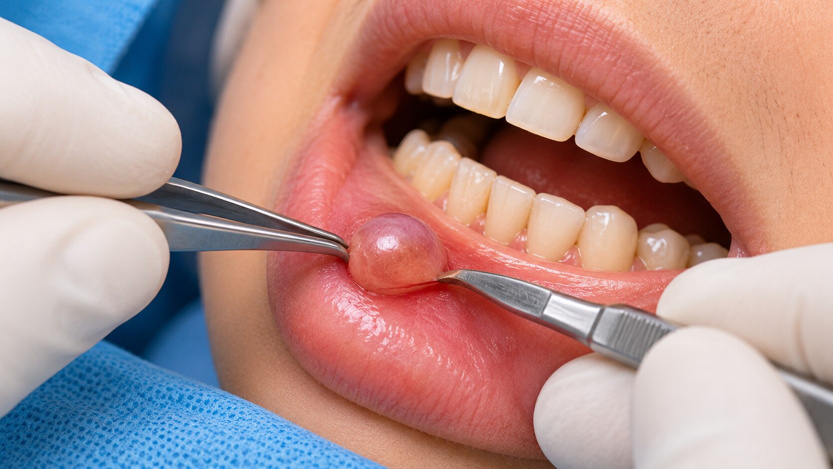
| Enucleation | Removes the complete cyst lining in one operation | Offers definitive treatment, but a large cavity can cause bleeding, nerve injury, tooth damage, or rarely mandibular fracture. |
| Marsupialisation | Opens the cyst into the mouth so it drains and shrinks | Reduces pressure on teeth, nerves, and sinus, but requires cleaning, reviews, and later removal of the remaining lining. |
| Decompression | Uses a tube or small opening to gradually reduce cyst size | Usually limits immediate bone removal, but treatment lasts longer and a second operation may be needed. |
| Partial jaw removal | Removes the cyst with surrounding jawbone | Reduces residual disease risk in selected aggressive lesions, but causes the greatest functional, nerve, tooth, fracture, and reconstruction risks. |
Upper-jaw surgery can create an opening into the maxillary sinus or nose, causing fluid passage and delayed healing. Posterior mandibular surgery can cause temporary or persistent numbness. Ask that the entire specimen undergo histopathology: a presumed cyst can be a tumour, and keratocysts recur more often than many other cysts.
These points belong in any discussion of oral cyst surgery risks mumbai.
What recovery should look like during the first week
During the first week, pain should gradually improve, swelling often peaks at 48–72 hours, and light blood-stained saliva can occur after oral cyst surgery. A white or pale-yellow coating over the socket is usually healing tissue, not pus. Sutures may feel tight or loosen as the wound closes.
| Symptom | Expected change | Contact the operating team urgently when |
|---|---|---|
| Pain | Moderate soreness that responds to prescribed medicine and improves daily | Pain worsens after initial improvement or becomes severe |
| Swelling or bruising | Swelling around the cheek, often greatest by day two or three, then settling | Swelling increases after the initial period, or fever and pus appear |
| Bleeding | Slow oozing controlled by firm gauze pressure | Bleeding continues despite sustained pressure or makes you feel faint |
| Numbness | Temporary altered feeling near the lower lip, chin, tongue, or cheek, especially after posterior-mandible surgery | Numbness is new, worsening, or persists beyond the review plan |
| Wound or sinus symptoms | Tenderness and a changing socket as it heals | Fluid passes between the mouth and nose, breathing or swallowing becomes difficult, or the wound opens widely |
Use prescribed medicines exactly as directed, keep the gauze in place as instructed, and choose soft, lukewarm foods. Avoid smoking, alcohol, vigorous rinsing, spitting, and strenuous exercise during the early healing period; clean the remaining teeth gently and use any prescribed rinse correctly. Attend dressing, drain, and wound reviews.
Tell the surgeon before treatment about diabetes, smoking, and poor oral hygiene. These factors increase infection and delayed-healing risks among oral cyst operation problems. Contact the clinic rather than waiting for the next appointment when symptoms worsen.
Why follow-up continues after the wound heals
Good oral cyst removal follow up continues after the gum closes because bone healing takes longer, and symptoms cannot show whether abnormal tissue remains. The surgeon must review the histopathology report, not assume that a radiolucency was a simple cyst.
A follow-up plan usually includes:
- An early appointment to inspect the wound, remove sutures, and review a drain or decompression device. Missed dressing reviews can allow infection or blockage.
- A pathology review confirming the lesion type. The entire specimen should undergo histopathology; an unexpected tumour or other jaw disease requires a different treatment plan and referral.
- A dental assessment for tooth vitality, mobility, bite, and the condition of neighbouring roots. Nerve testing matters after surgery near the inferior alveolar or lingual nerve.
- Follow-up radiographs, such as a periapical or panoramic X-ray, to document bone fill and detect residual or recurrent disease. CBCT is useful when three-dimensional detail is needed, including after large lesions or complex jaw surgery.
- Longer-term reviews based on the cyst type and operation. Decompression needs repeated measurements and device checks, while jaw surgery needs assessment of bone strength, facial sensation, and function.
Blood tests are not a substitute for tissue diagnosis and are ordered when infection, bleeding risk, or a medical condition requires them. Ask for the written pathology result and the planned imaging interval before you leave the consultation.
Questions to ask at an oral cyst removal consultation in Borivali
Before agreeing to treatment, use an oral cyst removal consultation in Borivali to obtain specific answers, not only a fee and surgery date.
1. What is the working diagnosis, and what evidence supports it? Ask whether the examination, periapical or panoramic X-ray, and CBCT show the lesion’s attachment to a tooth, its size, and its relationship to the inferior alveolar nerve or sinus.
2. Who will perform the operation? Confirm the surgeon’s oral and maxillofacial surgery training and whether they personally handle the planned procedure.
3. Which options apply: extraction with cyst removal, apicoectomy, enucleation, marsupialisation, decompression, or removal of part of the jaw? Ask why the recommended option offers a better balance of healing, recurrence, nerve injury, and jaw strength.
4. Can the tooth be preserved? Ask about root-canal treatment, tooth vitality testing, possible loosening, and the conditions that would require extraction. Do not assume an implant belongs in the same appointment; infection, bone healing, and the pathology result affect timing.
5. Will the entire specimen be sent for histopathology? A radiographic “cyst” can be another lesion, including a tumour.
6. What is the emergency pathway after hours? Get a phone number, hospital location, and instructions for uncontrolled bleeding, fever, worsening swelling, or breathing difficulty.
7. What are the review dates? Confirm wound checks, pathology review, nerve and tooth-vitality assessment, and follow-up radiographs to verify bone fill or detect recurrence. Dr. Karan Mehta's Mouth Jaw Face & Hair Transplant should explain these points before treatment.
Related services
Face A cyst is a water-filled cavity. The most common cyst in the oral region is a Radicular cyst arising at the root of teeth due to infection in it. View service → |
Jaw Jaw Cyst and Tumors Various cysts and tumors can arise in both upper jaw bone(maxilla) and lower jaw bone(mandible). View service → |
Frequently asked questions
When does a suspected oral cyst need urgent assessment?
Seek prompt assessment for rapidly increasing jaw swelling, fever, pus, difficulty swallowing or breathing, uncontrolled bleeding, or new facial numbness. Fluid between the mouth and nose, worsening sinus symptoms, or swelling near the eye also needs urgent review.
How does the operation change oral cyst removal risks?
Risk depends on the cyst’s size and location, its relationship to teeth and nerves, and whether it reaches the maxillary sinus. Ask which structures are nearby and whether removal requires tooth extraction, bone repair, or sinus management.
What should recovery look like during the first week?
Follow the surgeon’s instructions for cleaning, eating, medicines, and activity. Contact the clinic if pain or swelling worsens instead of improving, or if you develop fever, pus, bleeding, breathing difficulty, or new numbness.
Why is follow-up needed after the wound heals?
Follow-up confirms healing, reviews the laboratory pathology report, checks nearby teeth and bone, and identifies recurrence or another lesion that needs monitoring.
What should you ask at an oral cyst removal consultation in Borivali?
Ask what the lesion could be, which scans or tests are needed, whether a biopsy will be sent for pathology, which structures face risk, what recovery involves, and when follow-up is scheduled.
Keywords